
Table of Contents >> Show >> Hide
- What Is an X-Ray?
- Purpose of an X-Ray: Why Doctors Order It
- Types of X-Ray Exams
- How to Prepare for an X-Ray
- X-Ray Procedure: What Happens During the Test?
- After the X-Ray: Results and Follow-Up
- Possible Risks of an X-Ray
- How to Reduce Unnecessary X-Ray Exposure
- X-Ray vs. CT Scan vs. MRI vs. Ultrasound
- When Should You Call a Doctor After an X-Ray?
- Real-Life Examples of Why X-Rays Matter
- Experiences Related to X-Rays: What Patients Often Notice
- Conclusion
An X-ray is one of those medical tests that sounds futuristic, feels surprisingly ordinary, and often takes less time than finding a parking spot at the clinic. It is a quick imaging exam that uses a small amount of ionizing radiation to create pictures of structures inside the body, especially bones, teeth, the chest, and certain areas of the abdomen. In plain English: it helps doctors look under the hood without opening the hood.
Healthcare providers use X-rays to diagnose injuries, check symptoms, guide treatment, and monitor certain conditions. A broken wrist after a dramatic encounter with a skateboard? X-ray. A lingering cough that refuses to leave like an unwanted houseguest? Chest X-ray. A child who may have swallowed a small object? Yes, often an X-ray too.
Although the word “radiation” can make people nervous, most routine X-ray exams use a low dose. The key is appropriate use: an X-ray should be done when the medical benefit outweighs the possible risk. This guide explains the purpose of X-rays, how the procedure works, how to prepare, what results may show, and what possible risks patients should understand before the exam.
What Is an X-Ray?
An X-ray is a type of medical imaging test that sends controlled X-ray beams through the body. Different tissues absorb those beams in different ways. Dense materials, such as bone and metal, absorb more radiation and usually appear white on the image. Air, such as air inside the lungs, appears black. Fat, muscle, organs, and other soft tissues appear in shades of gray.
The image produced by a standard X-ray is called a radiograph. Today, most X-ray images are digital, which means they can be viewed on a computer screen, stored in electronic medical records, and shared with the healthcare team when needed. A radiologic technologist usually performs the exam, while a radiologista doctor trained in interpreting medical imagesreviews the images and sends a report to the ordering provider.
Purpose of an X-Ray: Why Doctors Order It
The main purpose of an X-ray is to help diagnose or monitor a medical problem. It is often chosen because it is fast, widely available, relatively inexpensive compared with many advanced imaging tests, and excellent for showing bones and certain chest conditions.
1. Diagnosing Broken Bones and Joint Problems
One of the most common uses of an X-ray is checking for fractures. If someone falls, twists an ankle, injures a shoulder, or has pain after a car accident, an X-ray can help reveal whether a bone is broken, displaced, or healing correctly.
X-rays can also show joint alignment, arthritis-related changes, bone spurs, and some signs of infection or tumors in bone. While an X-ray may not show every soft-tissue injury, such as a torn ligament, it is often the first step in figuring out what happened.
2. Evaluating Chest Symptoms
A chest X-ray can help doctors look at the lungs, heart size, ribs, and surrounding structures. It may be ordered for symptoms such as cough, shortness of breath, chest pain, fever, or suspected pneumonia. Chest X-rays can also help detect fluid around the lungs, collapsed lung, certain infections, and changes related to chronic lung or heart conditions.
Because chest X-rays are quick, they are especially useful in urgent care and emergency settings. They are not magic crystal balls, but they are very good at giving doctors an important first look.
3. Finding Swallowed Objects or Foreign Bodies
Children are curious. Sometimes that curiosity involves coins, toy pieces, batteries, or other objects that absolutely were not on the snack menu. X-rays may help locate swallowed or lodged objects, depending on what the object is made of. Metal objects show up well; some plastic or organic materials may be harder to see.
4. Checking Dental and Jaw Problems
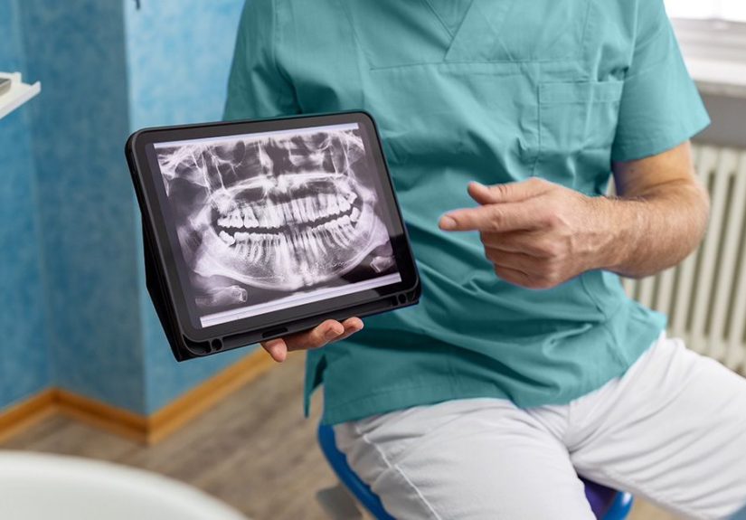
Dental X-rays help dentists see tooth decay, bone loss, impacted teeth, abscesses, and jaw structure. They are commonly used during routine dental care and before procedures such as wisdom tooth removal, root canal treatment, or dental implant planning.
5. Looking at the Abdomen
Abdominal X-rays may be used to evaluate certain digestive tract problems, bowel blockage, kidney stones that contain calcium, or swallowed objects. Some digestive X-ray exams use contrast material, such as barium, to make the esophagus, stomach, intestines, or colon easier to see.
6. Guiding Certain Medical Procedures
Some procedures use live or repeated X-ray imaging, called fluoroscopy, to guide instruments inside the body. This is different from a simple still-image X-ray. Fluoroscopy may be used for certain orthopedic, vascular, gastrointestinal, or pain-management procedures. Because fluoroscopy may involve more radiation than a single standard X-ray, the care team carefully balances image quality, procedure time, and safety.
Types of X-Ray Exams
There are many kinds of X-ray exams, each designed for a specific body area or medical question. Common examples include:
- Bone X-ray: Used for fractures, joint alignment, arthritis, and bone abnormalities.
- Chest X-ray: Used to evaluate lungs, heart size, ribs, and chest symptoms.
- Dental X-ray: Used to check teeth, roots, jawbone, and hidden decay.
- Abdominal X-ray: Used for certain digestive, urinary, or foreign-object concerns.
- Mammography: A specialized breast X-ray used for breast cancer screening or diagnosis.
- Fluoroscopy: A moving X-ray image used during certain procedures or contrast studies.
- CT scan: A more advanced test that uses X-ray technology and computer processing to create cross-sectional images.
Not every X-ray test has the same dose, purpose, or preparation. A single hand X-ray is very different from a CT scan or a long fluoroscopy-guided procedure. That is why patients should ask what exam is being done and why it is necessary.
How to Prepare for an X-Ray
Preparation depends on the body part being examined. For many basic X-rays, little or no preparation is needed. You may simply arrive, check in, answer a few safety questions, and be called back for the scan.
Before the Exam
Tell your healthcare provider and the X-ray technologist if you are pregnant, might be pregnant, or are breastfeeding. This is especially important for exams involving the abdomen, pelvis, lower back, or hips. In many cases, the risk from a medically necessary X-ray during pregnancy is small, but the care team may adjust the exam, use shielding when appropriate, or choose another imaging method such as ultrasound.
You should also mention recent X-rays or imaging tests, especially if you have had several exams in a short time. Keeping a personal list of imaging tests can help reduce unnecessary repeat imaging.
What to Wear
Wear comfortable clothing without metal snaps, zippers, hooks, or decorative hardware near the area being imaged. Metal can block the X-ray beam and create artifacts on the image. Depending on the exam, you may be asked to change into a gown.
Remove jewelry, eyeglasses, belts, hairpins, removable dental appliances, or body piercings if they are near the area being scanned. Think of it as temporarily de-accessorizing for science.
If Contrast Material Is Used
Some X-ray exams use contrast material to make certain organs or body spaces easier to see. Barium may be swallowed or given as an enema for digestive tract imaging. Iodine-based contrast may be injected for some procedures. If contrast is involved, tell your provider about allergies, kidney disease, asthma, diabetes, previous reactions to contrast, and all medications you take.
For some contrast exams, you may be asked not to eat or drink for a certain period beforehand. Always follow the specific instructions from your imaging center or healthcare provider.
X-Ray Procedure: What Happens During the Test?
The X-ray procedure is usually straightforward. A technologist positions your body so the correct area is lined up with the X-ray machine and image detector. You may sit, stand, or lie on a table depending on the exam.
Positioning
The technologist may take images from different angles. For example, a wrist injury may require front, side, and angled views. This helps the radiologist see the area clearly and avoid missing subtle fractures or alignment problems.
Holding Still
You will need to stay still while the image is taken. Movement can blur the picture, just like taking a shaky photo with a phone. For a chest X-ray, you may be asked to take a deep breath and hold it for a second or two. The actual image capture is usually very quick.
Will It Hurt?
The X-ray itself does not hurt. You do not feel the radiation. However, positioning may be uncomfortable if the injured area is painful. For example, moving a sore shoulder or swollen ankle into the correct position may feel unpleasant for a moment. The technologist will try to position you safely while still getting useful images.
How Long Does an X-Ray Take?
Many routine X-ray exams take only a few minutes once you are in the imaging room. More complex exams, multiple views, or contrast studies can take longer. Emergency results may be available quickly, while routine results may take hours to a couple of days depending on the facility and situation.
After the X-Ray: Results and Follow-Up
After the images are taken, the technologist checks whether they are clear enough. You may be asked to wait briefly in case another image is needed. Once the exam is complete, you can usually return to normal activities right away unless your provider gives different instructions.
A radiologist reviews the images and creates a report. Your primary doctor, emergency clinician, dentist, specialist, or other ordering provider explains the results and next steps. If the X-ray shows a fracture, pneumonia, bowel blockage, abnormal mass, or another concern, you may need treatment, additional imaging, lab tests, or referral to a specialist.
Possible Risks of an X-Ray
X-rays are generally considered safe when medically necessary and performed properly. Still, they use ionizing radiation, so it is reasonable to understand the possible risks.
Radiation Exposure
The main risk of an X-ray is exposure to a small amount of ionizing radiation. Ionizing radiation can damage DNA at high levels or after repeated exposures, and this may slightly increase the lifetime risk of cancer. However, most routine diagnostic X-rays use low doses, and the benefit of finding or ruling out a medical problem usually outweighs the risk.
The dose depends on the type of exam. A dental X-ray or hand X-ray uses a very small amount. A CT scan generally uses more because it takes many images from different angles. Fluoroscopy dose varies and can be higher during longer, complex procedures.
Pregnancy Considerations
If you are pregnant or may be pregnant, tell the healthcare team before the exam. Many medically needed X-rays can still be performed safely, especially when the area being imaged is away from the abdomen and pelvis. However, your provider may postpone the exam, use another test, or take extra precautions depending on the situation.
The goal is not to panic. The goal is to make a smart medical decision with the right information.
Children and X-Rays
Children are more sensitive to radiation than adults because their bodies are still developing and they have more years ahead for any potential effects to appear. Pediatric imaging should use child-sized settings and the lowest radiation dose needed to create a useful image. Parents can ask whether the facility adjusts X-ray settings for children.
Contrast Reactions
Most basic X-rays do not require contrast material. When contrast is used, possible side effects depend on the type. Barium may cause constipation or temporary stool color changes. Iodine-based contrast can sometimes cause warmth, nausea, itching, hives, or allergic-like reactions. Severe reactions are rare, but patients should always report prior contrast reactions.
False Reassurance or Missed Findings
No imaging test is perfect. A small fracture, early infection, soft-tissue injury, or subtle abnormality may not always appear on a standard X-ray. If symptoms continue despite a “normal” result, follow up with your healthcare provider. Sometimes repeat imaging, MRI, CT, ultrasound, or lab testing is needed.
How to Reduce Unnecessary X-Ray Exposure
You do not need to fear X-rays, but you also do not need extra ones for fun. This is medicine, not a photo booth.
- Ask why the X-ray is needed. A good question is, “How will this result change my care?”
- Share your imaging history. Mention recent X-rays, CT scans, or dental imaging.
- Tell the team about pregnancy. This helps them choose the safest approach.
- Use pediatric facilities when possible for children. Child-sized settings matter.
- Follow positioning instructions. Holding still can prevent blurry images and repeat scans.
- Keep copies of important imaging reports. This may help avoid duplicate testing.
X-Ray vs. CT Scan vs. MRI vs. Ultrasound
Different imaging tests answer different questions. An X-ray is excellent for bones and many chest concerns. A CT scan uses X-ray technology but provides more detailed cross-sectional images, which can help evaluate internal injuries, complex fractures, tumors, blood vessels, and many urgent conditions.
MRI uses magnetic fields and radio waves, not ionizing radiation. It is often better for soft tissues such as ligaments, brain structures, spinal discs, and certain joints. Ultrasound uses sound waves and is commonly used for pregnancy, gallbladder problems, pelvic organs, blood flow, and soft-tissue evaluation.
The “best” test depends on the medical question. More advanced does not always mean better. Sometimes the humble X-ray is exactly what is needed.
When Should You Call a Doctor After an X-Ray?
Call your healthcare provider if your symptoms get worse, your pain does not improve, you develop fever, swelling increases, breathing becomes difficult, or you do not receive results within the expected time. Also call if you had contrast material and develop symptoms such as hives, trouble breathing, severe dizziness, or persistent vomiting.
If you have severe chest pain, sudden shortness of breath, signs of stroke, major trauma, or another emergency, seek emergency care immediately.
Real-Life Examples of Why X-Rays Matter
The “It’s Probably Just a Sprain” Ankle
A person rolls an ankle during a weekend basketball game. It swells quickly, walking hurts, and everyone nearby becomes an unofficial sports medicine expert. An X-ray can help determine whether it is a sprain, fracture, or joint alignment problem. That difference matters because treatment may range from rest and support to immobilization or orthopedic care.
The Cough That Would Not Quit
A patient has fever, cough, and fatigue. A chest X-ray may help identify pneumonia or another lung issue. The result can guide treatment and help the provider decide whether antibiotics, further testing, or follow-up imaging is needed.
The Mystery Coin
A child swallows a coin. The parent is calm on the outside and absolutely not calm on the inside. An X-ray can help locate the coin and show whether it has moved into the stomach or is stuck somewhere that needs urgent attention.
Experiences Related to X-Rays: What Patients Often Notice
For many people, the most memorable part of an X-ray is not the machine itself. It is the small human moments around it: the paperwork, the chilly room, the awkward position, the technologist saying, “Hold still,” and the patient trying not to blink even though blinking has nothing to do with the ankle being scanned.
One common experience is surprise at how quick the test is. People may imagine a dramatic, high-tech process with buzzing equipment and suspenseful music. In reality, a routine X-ray is usually calm and efficient. The technologist positions you, steps behind a protective barrier, takes the image, and may reposition you for another view. The whole thing can feel almost too simple for something that provides such useful information.
Another common experience is mild discomfort during positioning. The X-ray beam does not hurt, but if you have a painful injury, moving into the correct angle can be uncomfortable. For example, someone with a wrist fracture may need to turn the hand slightly, or a patient with knee pain may need to straighten the leg. Technologists are trained to help patients get the needed image while minimizing discomfort, but a few seconds of “please let this be over” is not unusual.
Patients also often notice the importance of clear instructions. When the technologist says, “Take a deep breath and hold it,” during a chest X-ray, that breath helps expand the lungs and improve the image. When they say, “Do not move,” they are not being dramatic; motion can blur the image and may require another exposure. In this tiny contest between you and the blur, stillness wins.
Parents may have a different emotional experience when their child needs an X-ray. Even when the test is low risk, seeing a child in a medical setting can be stressful. The best approach is usually calm preparation: explain that the camera takes a picture of the inside of the body, it does not touch or pinch, and staying still helps the picture come out clearly. For younger children, a favorite stuffed animal, a simple explanation, or a calm adult voice can make the exam smoother.
Pregnant patients may feel extra concern, and that is understandable. The most helpful step is to tell the care team immediately. In many cases, the provider can explain whether the exam is necessary, whether another test could work, and what precautions may be used. The conversation should be practical, not scary. Avoiding a needed diagnosis can sometimes be riskier than the imaging itself.
People who receive contrast material may remember different sensations. With some injected contrast agents, patients report warmth or a strange taste. With barium studies, the drink may taste chalky or heavy. These experiences are usually temporary, but patients should report allergies, prior contrast reactions, kidney problems, or unusual symptoms after the test.
Another real-world issue is waiting for results. In emergency settings, results may come quickly. In routine outpatient settings, it may take longer for the radiologist’s report to reach the ordering provider. Patients sometimes see image files before they understand the report, which can lead to unnecessary worry. A cloudy-looking area or unfamiliar phrase is not always a disaster. Medical images need expert interpretation and clinical context.
Finally, many patients learn that an X-ray is one piece of the puzzle, not the entire puzzle. A normal X-ray can be reassuring, but it does not always explain pain, swelling, or other symptoms. Soft-tissue injuries, early stress fractures, or certain internal conditions may require follow-up, repeat imaging, or a different test. The best experience comes from asking questions, understanding the reason for the exam, and following up if symptoms do not match the results.
Conclusion
An X-ray is a fast, useful, and widely available imaging test that helps healthcare providers diagnose injuries, evaluate symptoms, guide treatment, and monitor certain conditions. It is especially valuable for bones, chest problems, dental concerns, swallowed objects, and many urgent medical questions.
Like any medical test, an X-ray should be used thoughtfully. Most routine X-rays involve low radiation exposure, and the benefit of an accurate diagnosis usually outweighs the small potential risk. Patients can help by sharing pregnancy status, recent imaging history, allergies, and medical conditions. They can also ask why the exam is needed and how the results will guide care.
The bottom line: an X-ray is not something to fear, but it is something to understand. When ordered for the right reason, performed with proper technique, and interpreted by trained professionals, it can be a powerful tool for getting answers quicklyand sometimes, getting you back on your feet with less guessing and more confidence.