Table of Contents >> Show >> Hide
- What Are Dog Eyelid Growths?
- Why Eyelid Growths Matter (Even When They’re Benign)
- Symptoms of a Dog Eyelid Growth
- What Causes Eyelid Growths in Dogs?
- How Veterinarians Diagnose Dog Eyelid Growths
- Treatment Options for Dog Eyelid Growths
- Dog Eyelid Growth Surgery: What to Expect
- Recovery After Dog Eyelid Growth Treatment
- When Observation Is Reasonable vs. When Removal Is Better
- Prevention and Monitoring Tips for Dog Owners
- Final Takeaway
- Extended Experience Section (Approx. 500+ Words): Real-World Recovery Patterns Owners Often See
- Conclusion SEO Tags (JSON)
If you’ve ever looked at your dog and thought, “Was that little bump on the eyelid there yesterday?” you are definitely not alone. Eyelid growths in dogs are common, especially in older dogs, and while many are benign (the “annoying but manageable” kind), they still matter because the eyelid is prime real estate. Even a tiny growth can rub the cornea, trigger irritation, and turn a small problem into a squinty, teary, uncomfortable eye.
The good news: many dog eyelid growths can be diagnosed and treated successfully, especially when caught early. The trick is not guessing at home. A bump that looks harmless can be inflammatory, infectious, or neoplastic (tumor-related), and the treatment plan depends on what it actually is.
This guide walks you through what dog eyelid growths are, how veterinarians diagnose them, what treatment options are commonly used, and what recovery usually looks like. We’ll also cover warning signs, practical post-op tips, and a bonus experience section at the end so you know what this often looks like in real life.
What Are Dog Eyelid Growths?
“Dog eyelid growths” is a broad term. It can describe small bumps on the eyelid margin, masses inside the lid, or growths near the eye that affect blinking and tear film distribution. Some are true tumors, some are gland-related swellings, and some are inflammatory lesions that imitate tumors like they’re auditioning for a medical drama.
Common types of eyelid growths in dogs
- Meibomian gland adenoma (very common, usually benign)
- Meibomian gland epithelioma / hyperplasia
- Papilloma (wart-like growth)
- Melanocytic growths (pigmented lesions; behavior varies)
- Histiocytoma (more common in younger dogs, can self-resolve in some cases)
- Mast cell tumor (requires more careful workup)
- Chalazion / lipogranulomatous inflammation (blocked or ruptured meibomian gland swelling)
- Inflammatory blepharitis lesions (can mimic a mass)
In dogs, many eyelid tumors are benign, and meibomian gland tumors are among the most common. That said, “common” is not the same as “ignore it.” A benign mass can still be painful if it scratches the cornea or disrupts blinking.
Why Eyelid Growths Matter (Even When They’re Benign)
The eyelids are not just little face curtains. They protect the eye, spread tears, and help keep the cornea smooth and comfortable. When a growth forms on the lid margin, it can rub the cornea like a tiny rock in a shoe. Your dog may respond with squinting, tearing, pawing, and eye discharge.
Left untreated, some growths can cause repeated irritation, corneal scratches, ulcers, secondary infection, and chronic inflammation. In other cases, the concern is not friction but cancer risk or local tissue invasion. That’s why veterinary evaluation matters early.
Symptoms of a Dog Eyelid Growth
Some dogs act completely normal at first, especially if the mass is small. Others make it obvious by turning into a one-eye pirate with dramatic squinting.
Common signs to watch for
- A visible bump, wart-like growth, or pigmented mass on the eyelid
- Squinting or blinking more than usual
- Tearing or mucus discharge
- Redness of the eye or eyelid
- Pawing or rubbing the eye
- Bleeding, ulceration, or crusting on the mass
- Change in the way the eyelid opens/closes
- Cloudiness of the eye (possible corneal irritation or ulcer)
Signs that need urgent veterinary attention
- Sudden severe squinting or the eye won’t open
- Blue/white haze on the cornea
- Bleeding or rapid growth
- Marked swelling, pain, or repeated rubbing
- Vision changes or obvious distress
What Causes Eyelid Growths in Dogs?
There is no single cause for most eyelid tumors. Like many tumors, they likely result from a mix of age, genetics, cell changes, and environmental factors. Some lesions are not tumors at all and come from blocked glands, inflammation, or infection.
Age is a major factor for many eyelid masses, especially meibomian gland tumors. Certain malignant skin-related tumors may be associated with UV exposure in susceptible animals, but in day-to-day practice, the most useful question is less “why did this happen?” and more “what exactly is it?”
How Veterinarians Diagnose Dog Eyelid Growths
Diagnosis starts with a physical and eye exam, but the real answer often comes from sampling or removing the tissue. Eyelid masses are one of those situations where “looks like” is helpful, but “pathology says” is better.
Step 1: Eye exam and history
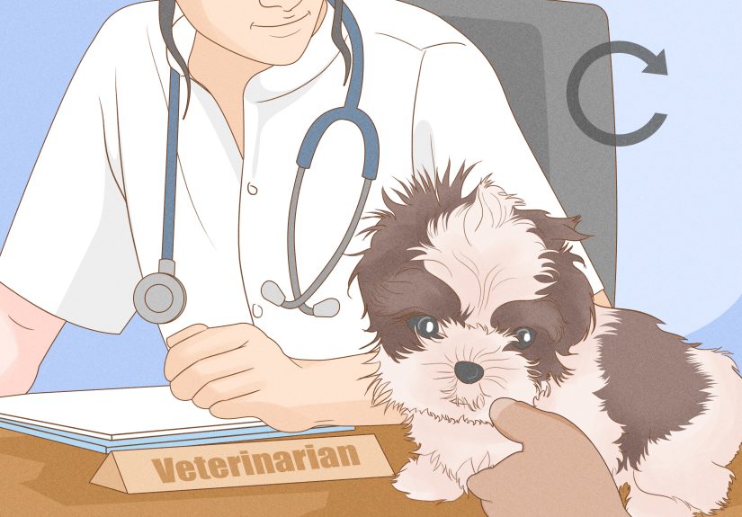
Your veterinarian will assess size, location, color, rate of growth, and whether the mass touches the cornea. They will also check for ocular surface damage and signs of infection or ulceration. If needed, you may be referred to a veterinary ophthalmologist, especially for tricky locations, larger masses, recurrent lesions, or suspected malignancy.
Step 2: Diagnostic tests (when indicated)
- Fluorescein stain to check for corneal ulcers
- Tear testing if dry eye is suspected
- Cytology / FNA (fine-needle aspirate) for certain masses
- Punch biopsy for atypical or suspicious lesions
- Histopathology (microscopic exam of removed tissue) for definitive diagnosis
- Staging tests (bloodwork, lymph node assessment, chest imaging) if malignancy is suspected
In many cases, especially when a mass is small but irritating, the growth is removed and submitted for histopathology. This gives the most reliable diagnosis and helps guide prognosis and follow-up.
Treatment Options for Dog Eyelid Growths
Treatment depends on the size, location, type of growth, and whether it’s irritating the eye. There is no one-size-fits-all approach. Tiny masses may be monitored, but growing or rubbing lesions are usually treated.
1) Monitoring (watchful waiting)
In select cases, especially tiny, slow-growing masses with no corneal contact and no irritation, your veterinarian may recommend monitoring. Lubricating eye medication may be used to protect the cornea if there is mild friction. Monitoring is not “doing nothing”; it means tracking size, appearance, and symptoms carefully.
2) Debulking + cryotherapy (freezing therapy)
For many small eyelid margin masses (especially common meibomian-related growths), debulking the visible portion and treating the area with cryotherapy is a common option. Cryotherapy helps destroy residual cells left behind after trimming. It can be very effective, but regrowth can still happen, and temporary swelling, crusting, skin irritation, or pigment changes may occur during healing.
3) Surgical excision (wedge or pentagonal resection / V-plasty)
Larger masses, recurrent lesions, or masses with a broader base often require formal surgical removal with eyelid reconstruction. This may involve a wedge or pentagonal excision (often described in practice as V-plasty for certain cases). The goal is to remove the mass while preserving eyelid function and smooth margin alignment.
Eyelid surgery is delicate because even a tiny mismatch in the lid margin can irritate the cornea long-term. This is why a veterinary ophthalmologist is often recommended for complicated lesions or lesions affecting a large portion of the eyelid.
4) Laser-assisted removal (selected cases)
Some practices use CO2 laser techniques for small eyelid masses. The exact method depends on the mass type, surgeon preference, and available equipment.
5) Medical management (for inflammatory or non-neoplastic lesions)
Not every bump needs surgery. Some eyelid swellings are inflammatory, infected, or gland-blockage-related and may be managed with topical medications, anti-inflammatory treatment, or other supportive care. This is one more reason not to self-diagnose from a photo.
6) More advanced surgery / oncology care
If the mass is suspected or confirmed to be malignant, treatment may require biopsy first, wider surgical margins, reconstructive techniques, and sometimes additional diagnostics or therapies. The plan is individualized based on tumor behavior and whether the lesion has spread.
Dog Eyelid Growth Surgery: What to Expect
Before surgery
- Exam to assess the mass and corneal health
- Discussion of surgical approach and anesthesia/sedation plan
- Potential bloodwork (especially in older dogs)
- Possible referral to ophthalmology for complex cases
Day of procedure
Depending on the size and technique, your dog may have brief anesthesia, sedation, or, in selected calm patients and specialized settings, local anesthesia approaches. Many eyelid procedures are outpatient, though some specialty hospitals may keep pets longer depending on the surgery and the patient’s needs.
After surgery
Removed tissue is commonly sent for histopathology. This is the part where the pathologist settles the “what is it?” debate once and for all.
Recovery After Dog Eyelid Growth Treatment
Recovery varies by procedure, but most dogs do well when owners follow post-op instructions closely. The two biggest recovery themes are: protect the eye and don’t stop meds early just because it looks better.
Typical recovery timeline (general)
- Days 1–3: swelling, redness, squinting, and mild discharge are common
- Days 3–7: swelling may peak (especially after cryotherapy), then improve
- Week 1–2: tissue healing progresses; ulceration/crusting from cryotherapy may resolve
- About 10–14 days: many dogs are rechecked; sutures may be removed if non-absorbable
- Longer-term: monitor for regrowth, irritation, or lid margin changes
Common post-op instructions
- E-collar (cone): extremely important after eye surgery
- Topical medications: antibiotics, lubricants, or anti-inflammatory drops/ointment as prescribed
- Pain medication: use exactly as directed
- Activity restriction: avoid rough play and face rubbing
- Recheck visits: keep them, even if the eye looks “basically fine”
Eye surgeries can look dramatic during the first week, especially if cryotherapy was used. Temporary swelling, edema, and even pigment changes can happen. What matters most is steady improvement and good comfort.
Potential complications during recovery
- Corneal ulceration
- Infection or increased inflammation
- Suture irritation
- Eyelid margin misalignment
- Tumor regrowth / recurrence
- Abnormal eyelash direction or chronic corneal irritation
If your dog becomes more painful, more squinty, or the eye gets cloudier instead of better, contact your vet promptly. Eye problems are not great candidates for a “let’s give it a few more days” experiment.
When Observation Is Reasonable vs. When Removal Is Better
Observation may be reasonable if:
- The mass is very small
- It is not touching the cornea
- There is no redness, discharge, or squinting
- The dog is a high anesthesia risk
- Your vet has examined it and recommended monitoring
Removal is often preferred if:
- The mass is growing
- It rubs the cornea or disrupts blinking
- There is recurrent irritation, discharge, or ulceration
- The lesion looks atypical or suspicious for malignancy
- It has recurred after prior treatment
A practical rule of thumb: if the eyelid growth is making your dog uncomfortable, it usually deserves more than “we’ll just watch it.” Comfort and corneal protection matter a lot.
Prevention and Monitoring Tips for Dog Owners
- Check your dog’s eyes during grooming (especially senior dogs)
- Take a clear photo with the date if you notice a bump
- Note growth rate, discharge, squinting, and rubbing behavior
- Do not squeeze, cut, or medicate a growth without veterinary advice
- Use only prescribed eye medications (the wrong drops can worsen some conditions)
- Schedule follow-ups if your dog has had an eyelid mass removed before
Final Takeaway
Dog eyelid growths are common, and many are benign, but “benign” does not always mean “harmless.” Because the eyelid directly affects corneal protection and comfort, even a small growth can cause real problems. The best outcomes usually happen when a veterinarian evaluates the lesion early, protects the ocular surface, and confirms the diagnosis with cytology, biopsy, or histopathology when needed.
If your dog has a new eyelid bump, don’t panicbut don’t ignore it either. Early diagnosis, the right treatment plan, and good recovery care can make the difference between a quick fix and a long, squinty saga.
Extended Experience Section (Approx. 500+ Words): Real-World Recovery Patterns Owners Often See
The experiences below are composite examples based on common veterinary ophthalmology scenarios (not one specific dog), included to help you recognize what diagnosis, treatment, and recovery often feel like in real life.
Experience 1: “It looked tiny, but my dog wouldn’t stop blinking”
An older small-breed dog develops a 2–3 mm bump on the upper eyelid margin. At first, the owner thinks it’s just a skin tag because the dog is eating, playing, and still enthusiastically demanding snacks. Within a week, though, there’s more tearing and frequent blinking on that side. The primary vet stains the eye and finds mild corneal irritation from contact with the mass. Because the lesion is small and on the margin, the dog is referred for debulking plus cryotherapy. Recovery is a little messy-looking for the first few days: swelling, some crusting, and a deeply offended expression whenever the cone appears. But comfort improves quickly, and the recheck shows healing without corneal damage. Histopathology confirms a benign meibomian gland adenoma. The owner’s biggest takeaway: “I wish I had gone in sooner instead of waiting for it to get more annoying.”
Experience 2: “We watched it for months, then it started rubbing the eye”
A senior retriever has a slow-growing eyelid bump that was initially monitored because it was tiny, not touching the cornea, and not causing discharge. The vet recommended photos and monthly checks. For a while, that plan worked. Then the mass became broader and started changing how the eyelid sat against the eye. The dog developed intermittent redness and pawing, especially after long walks on windy days. At that point, observation was no longer the best plan. Surgical excision with eyelid reconstruction was chosen to preserve lid margin function. The owner reports the hardest part of recovery wasn’t the surgeryit was cone compliance. “My dog acted like the cone was a personal betrayal.” Still, by following the medication schedule and keeping the cone on, the incision healed well, and the dog returned to normal activity after the recheck. The key lesson here: monitoring can be appropriate, but the plan needs to change if comfort or corneal safety changes.
Experience 3: “It turned out not to be a tumor after all”
A middle-aged dog develops a firm eyelid swelling that looks suspiciously like a tumor. The owner assumes surgery is inevitable. During the exam, the veterinarian considers a chalazion or inflammatory lesion in the differential because the swelling pattern and tissue feel are a little unusual. After a diagnostic workup and targeted treatment, the lesion improves significantly without a full mass excision. This experience is a great reminder that not every eyelid lump is a cancerous or surgical problem. It also shows why internet image comparisons can mislead peoplemany different eyelid conditions can look similar in a cellphone photo.
Experience 4: “The pathology report changed the follow-up plan”
In another common scenario, a dog has an eyelid mass removed because it is recurrent and irritating. The surgery goes well, the eye looks much better within two weeks, and everyone expects the story to end there. Then the pathology report identifies a tumor type that warrants closer monitoring than a routine benign adenoma. The dog now gets regular rechecks, and the owner is taught what signs of recurrence to look for (new bump, redness, squinting, discharge, lid margin distortion). This experience highlights one of the most important points in eyelid mass care: how the growth looks is useful, but histopathology often determines the long-term plan. In other words, the lab report can be the plot twist that improves your dog’s outcome.
Across all of these experiences, the pattern is consistent: early evaluation, corneal protection, proper diagnosis, and strict recovery care lead to the best results. Also, yes, most dogs hate the coneuntil the eye stops hurting.