Table of Contents >> Show >> Hide
- Why the eye keeps showing up in Alzheimer’s conversations
- What researchers are looking for during eye testing
- So… what counts as a “routine” eye exam?
- Can an eye exam diagnose Alzheimer’s today?
- Where routine eye exams can help right now (even before “Alzheimer’s screening” exists)
- What to ask at your next eye appointment
- The future: “Oculomics,” AI, and smarter screening
- Experiences from the real world: what people notice (and what eye care can change)
- Conclusion: the eye exam isn’t a crystal ballbut it might become a useful flashlight
- SEO Tags
Imagine if your annual eye exam didn’t just answer the eternal question “Better one… or two?” but also whispered something useful about your brain health. That ideausing the eyes as a “window to the brain”is one of the most fascinating frontiers in Alzheimer’s research right now.
Here’s the careful truth: routine eye exams cannot diagnose Alzheimer’s disease today. But a growing body of research suggests that certain retinal and visual changes may track with cognitive decline or even appear before obvious memory symptoms. In the future, it’s possible that the same scans already used to monitor glaucoma and macular degeneration could help flag people who might benefit from earlier cognitive screening.
So, could routine eye exams help spot early signs of Alzheimer’s? Potentiallyeventuallyand in a “clues, not conclusions” kind of way. Let’s unpack what scientists are finding, what eye doctors can do right now, and what’s still firmly in the “promising but not ready” stage.
Why the eye keeps showing up in Alzheimer’s conversations
The retina isn’t just “part of the eye.” It’s actually nervous system tissue that processes light and sends signals to the brain through the optic nerve. The retina also has a dense network of tiny blood vesselsexactly the kind of microcirculation researchers care about when studying brain aging.
That makes the eye appealing for early detection research for three big reasons:
- Access: You can image the retina non-invasively in minutes.
- Detail: Modern scans can measure retinal layers down to microns (that’s “smaller than your patience in a DMV line”).
- Overlap: Alzheimer’s involves nerve cell changes, inflammation, and vascular changesthings that may leave footprints in retinal structure and blood flow.
Researchers sometimes call this broader trend “oculomics”: extracting meaningful health signals from eye data, often with the help of AI.
What researchers are looking for during eye testing
When headlines say “an eye test can detect Alzheimer’s,” they usually mean one of several research approachesmost of which build on tools already used in regular eye care.
1) Retinal nerve fiber layer thinning
One of the most studied findings is that people with Alzheimer’s (and sometimes mild cognitive impairment, or MCI) may have thinner retinal layersespecially the retinal nerve fiber layer (RNFL) and ganglion cell layers. These layers contain nerve fibers and cells that help transmit visual information.
Sounds straightforwarduntil you remember that glaucoma, normal aging, and several other conditions can also thin these layers. So thinning isn’t a fingerprint. It’s more like a smudge: interesting, but not uniquely identifying.
2) Retinal blood vessel changes (especially with OCT angiography)
Optical coherence tomography angiography (OCT-A) is a scan that maps retinal blood flow without dye. Some studies have found that people with Alzheimer’s or MCI show differences in retinal microvasculaturesuch as changes in vessel density or perfusion patterns.
A practical way to think about it: if the brain’s small vessels are struggling, the retina’s small vessels might show related stress signalsbecause they’re built from the same biological “plumbing” playbook.
3) Amyloid and tau signals in the eye (experimental)
The “dream scenario” is to detect hallmark Alzheimer’s proteinslike beta-amyloid and tauusing eye-based methods. There’s research exploring specialized imaging, fluorescent markers, and advanced analysis techniques (including retinal texture features and other “pattern” signals).
Important reality check: this is not part of routine eye care, and many approaches are still being validated. But it’s a major reason the eye has become such a hot topic in Alzheimer’s detection science.
4) Pupil response and eye-movement clues
Your pupils don’t just react to light; they also react to mental effort. Researchers study task-evoked pupillary responses (how the pupil changes during cognitive tasks) because they may reflect brain systems involved early in Alzheimer’s risk.
Eye movement patternslike how someone scans a face or readsare also being studied. Some people with early cognitive changes may show subtle differences in visual attention and tracking long before they say, “I’m worried about my memory.”
5) Visual function tests: contrast, color, depth, and motion
Alzheimer’s isn’t only memory. For some people, early issues involve visual processing: trouble with contrast sensitivity (seeing edges clearly), judging distance, distinguishing certain colors, or navigating in visually busy environments.
There’s also a less common Alzheimer’s-related condition called posterior cortical atrophy (PCA), where visual and spatial symptoms can show up earlythings like difficulty reading, judging distances, or recognizing objectssometimes before obvious memory problems.
So… what counts as a “routine” eye exam?
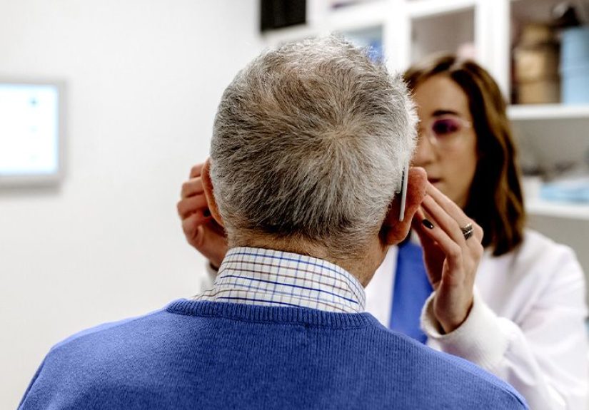
In real-life eye care, “routine” can range from a basic vision check to a full comprehensive eye exam with dilation and imaging. Here’s what many comprehensive exams may include:
- Visual acuity and refraction: glasses/contacts prescription
- Eye pressure measurement: screening for glaucoma risk
- Dilated retinal exam: direct look at the optic nerve and retina
- Retinal photos (fundus imaging): documentation of retinal appearance
- OCT: cross-sectional imaging of retinal layers (commonly used for glaucoma and macular disease)
- Sometimes OCT-A: a more specialized blood-flow map
Here’s the key point: many of the tools researchers use already exist in mainstream eye care. The missing piece is not the camerait’s the clinical proof that certain patterns reliably predict Alzheimer’s risk in a way that’s accurate, ethical, and useful.
Can an eye exam diagnose Alzheimer’s today?
No. Not on its own, and not as a standard medical practice in the U.S. right now.
What eye-related findings can do today is more like this:
- Raise questions when someone has unusual visual complaints or unexplained changes.
- Support research that may eventually create screening tools.
- Improve quality of life by treating correctable vision issues that can mimic or worsen confusion.
Why the caution? Because Alzheimer’s early detection is a high-stakes arena. A screening tool must be extremely reliableotherwise you end up with two bad outcomes:
- False positives: people panic, pursue expensive testing, or get labeled incorrectly.
- False negatives: people feel falsely reassured and delay care.
On top of that, eye findings are often not specific. For example, retinal thinning might reflect glaucoma, diabetes, high blood pressure, or just aging. That means researchers must prove that the “Alzheimer’s signal” remains meaningful after accounting for common eye and vascular conditions.
Where routine eye exams can help right now (even before “Alzheimer’s screening” exists)
1) Fixing vision may protect brain healthor at least remove a big obstacle
One of the strongest, most practical takeaways from recent research is that vision impairment is associated with dementiaand some estimates suggest a meaningful portion of dementia cases in older adults could be linked to vision problems, especially when considering distance vision, near vision, and contrast sensitivity together.
That does not prove that blurry vision causes dementia. But it does support a simple, helpful idea: keeping vision as strong as possible helps people stay active, social, and independentall things tied to better cognitive outcomes.
2) Eye care can reduce “cognitive load”
When vision is poor, the brain works harder to interpret the world. That extra effort can look like forgetfulness, irritability, withdrawal, or difficulty following conversationsespecially in older adults already dealing with stress, sleep issues, or medical complexity.
In other words: sometimes the “memory problem” is partly a “I literally can’t see the sign / face / pill label” problem. Treating cataracts, updating a prescription, managing glaucoma, or addressing dry eye won’t cure Alzheimer’sbut it can make daily life dramatically easier.
3) Eye doctors may be first to hear certain early complaints
People don’t always walk into a clinic saying, “Hello, I’m worried about my hippocampus.” They say things like:
- “Reading feels harder, but my glasses don’t seem to fix it.”
- “I’m having trouble driving at dusk or in rain.”
- “Busy patterns make me dizzy.”
- “I keep misjudging steps and curbs.”
Those symptoms can come from many causeseye disease, medication effects, vestibular issues, or ordinary aging. But they can also overlap with early changes in visual processing and attention. A thorough eye exam helps separate “eye problem” from “brain processing problem,” and that can guide next steps.
What to ask at your next eye appointment
If Alzheimer’s runs in your familyor you’re simply curiousyour eye visit is a reasonable place to start a broader health conversation. A few grounded questions:
- “Can you check my retina and optic nerve health thoroughly?” (This usually means a dilated exam and/or imaging.)
- “Do you use OCT, and would it be useful in my case?” (Especially if you have glaucoma risk, diabetes, or unexplained vision issues.)
- “Is my contrast sensitivity or visual function okay?” (Not every clinic measures this routinely, but some can.)
- “If my scan looks unusual, what would you recommend next?” (This can lead to practical referralsprimary care, neurology, or further eye testing.)
Tip: If you’re supporting a parent or grandparent, bring a short list of real-life exampleslike difficulty reading mail, getting lost in familiar stores, or frequent trips and falls. Specific observations beat vague worries every time.
The future: “Oculomics,” AI, and smarter screening
Many researchers are excited about a future where existing retinal images (fundus photos, OCT, OCT-A) can be analyzed with validated algorithms to identify patterns linked to Alzheimer’s pathology or dementia risk. The appeal is obvious: eye imaging is fast, relatively affordable, and already widely used.
But for this future to be helpfulnot chaoticseveral things must happen:
- Standardization: clinics and studies need consistent imaging methods and measurements.
- Validation: findings must hold up across diverse populations and real-world settings.
- Clinical pathways: if a scan flags “higher risk,” there must be clear next steps (and support).
- Ethics and consent: people deserve transparency about what is being screened and how results are used.
So yesroutine eye exams may eventually play a role in earlier Alzheimer’s detection. But the most responsible framing today is: eye exams may help identify risk signals that prompt further evaluation, rather than serving as a stand-alone diagnostic test.
Experiences from the real world: what people notice (and what eye care can change)
Research is exciting, but everyday experience is where this topic becomes personal. And in real clinics and homes, the “eye exam and Alzheimer’s” connection often shows up in a surprisingly practical way: visual confusion can look like cognitive declineand cognitive decline can make vision problems harder to manage.
Caregivers frequently describe a stage where a loved one insists, “My eyes are fine,” while also doing things that don’t match that claimmissing the chair when sitting down, stepping over a dark rug like it’s a hole, or abandoning reading because “the words move.” Sometimes the eye exam reveals a straightforward culprit: cataracts dulling contrast, an outdated prescription, or dry eye causing fluctuating blur. Fixing those doesn’t erase dementia, but families often report a noticeable change in confidenceless hesitation on stairs, fewer headaches, and fewer moments where frustration boils over because the world feels visually unreliable.
Eye clinics also see the flip side. A patient may have a perfectly reasonable new glasses prescription on paper, but they keep returning saying the glasses “don’t work.” When the clinician digs deeper, the complaint isn’t crispnessit’s processing: difficulty tracking lines of text, trouble finding items against a similar-colored background, or feeling overwhelmed by patterned carpet and crowded shelves. Those experiences can be a hint to consider broader screening, especially if the eye structures themselves look healthy.
Then there are the “in-between” experiencespeople who function well but quietly adapt. They stop driving at night, avoid unfamiliar routes, and choose restaurants with brighter lighting because dim menus feel impossible. It’s easy to chalk this up to normal aging (and sometimes it is). But in Alzheimer’s research contexts, subtle changes in contrast sensitivity, visual attention, or eye movements are exactly the kind of early signals scientists are studying. The important point for real life is not to self-diagnoseit’s to name the specific change and bring it to a clinician who can separate eye disease from brain-based visual processing changes.
Another common experience involves logistics: people with memory issues often struggle to follow eye-drop schedules, keep track of multiple pairs of glasses, or communicate symptoms accurately. Families sometimes discover that “noncompliance” is really a systems problemlabels too small to read, instructions too complex, or bifocals that are disorienting when spatial perception is already shaky. In these cases, an eye care team can help with practical adjustments: simplifying eyewear, improving contrast at home, recommending brighter task lighting, and coordinating with primary care when safety issues (like frequent falls) appear.
Finally, research volunteers often describe the emotional side of eye-based screening: the hope that a quick scan could offer clarity, mixed with the fear of learning something before they feel ready. That’s why the future of eye exams as Alzheimer’s screening must include not only better technology, but also better communicationclear explanations, informed consent, and a plan for what happens after a “flag.” Because an early clue is only helpful if it leads to support, not just worry.
Conclusion: the eye exam isn’t a crystal ballbut it might become a useful flashlight
Routine eye exams can’t diagnose Alzheimer’s today. Still, the science is moving in an intriguing direction: retinal structure, retinal blood flow, pupil responses, and visual function tests may provide early clues about brain health. The most useful action right now is also the least flashy: protect and correct vision. Clear vision supports independence, safety, and engagementthree things every brain appreciates.
So keep the annual eye exam on your calendar. If you ever needed an excuse, you can now say you’re not being vainyou’re being neurologically proactive.