
Table of Contents >> Show >> Hide
- Bronchitis 101: What’s actually inflamed?
- What a chest X-ray can (and can’t) “see”
- So how do X-rays help with bronchitis diagnosis?
- When doctors are more likely to order a chest X-ray
- How to interpret X-ray results in a bronchitis workup
- What the chest X-ray appointment is like
- If the X-ray is normal but you still feel awful
- X-rays vs other tests doctors may use
- Safety notes: radiation, repetition, and smart imaging
- When to seek urgent care (X-ray or not)
- Bottom line: what X-rays really do for bronchitis
- Real-Life Experiences: What patients (and clinicians) notice around chest X-rays for “bronchitis cough”
Bronchitis is one of those “I can’t stop coughing” problems that makes you question every life choice that led you to breathing air. And when you finally drag yourself to a clinic, you might hear: “Let’s get a chest X-ray.”
So… does an X-ray diagnose bronchitis?
Not exactly. But it can still be incredibly helpfulmostly because it helps your clinician figure out what your cough isn’t (like pneumonia), and whether anything more serious is going on. Think of a chest X-ray as a bouncer at a crowded party: it’s great at spotting the troublemakers, even if it doesn’t know everyone’s name.
Bronchitis 101: What’s actually inflamed?
Bronchitis means inflammation of the bronchial tubesthe airways that carry air in and out of your lungs. There are two main types:
Acute bronchitis
This is the common “bad cough after a cold” situation. It usually comes from a virus (like the ones that cause colds and flu), so antibiotics often won’t help. The cough can linger for weeks even after you feel mostly okaybecause your airways are irritated and healing.
Chronic bronchitis
This is long-term inflammation with a productive cough that lasts for months and tends to come back year after year. It’s often linked with smoking and chronic obstructive pulmonary disease (COPD). Chronic bronchitis is diagnosed based on symptoms/history and breathing tests (like spirometry), not just imaging.
What a chest X-ray can (and can’t) “see”
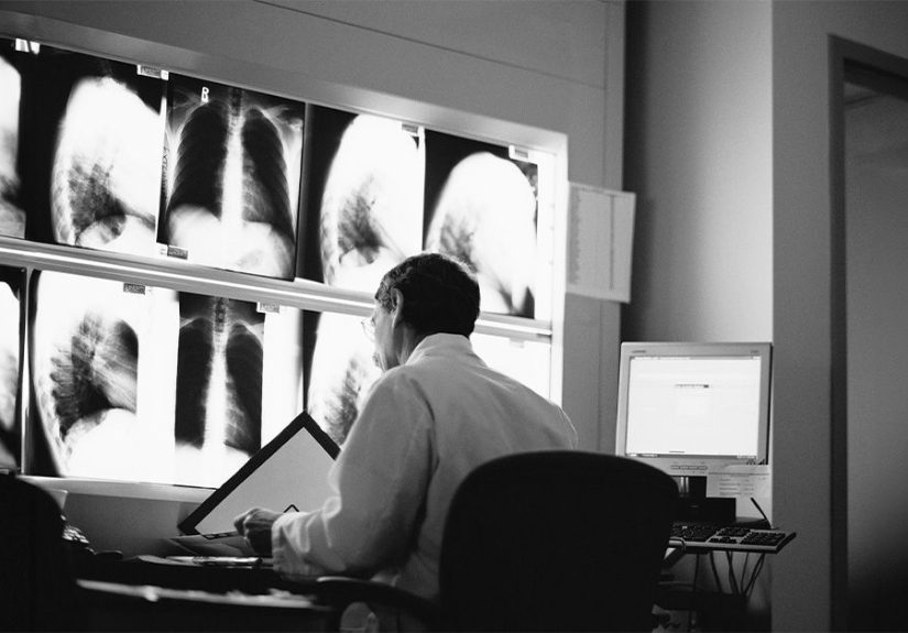
A chest X-ray (also called a chest radiograph) is a quick imaging test that creates pictures of your lungs, heart, and chest structures. It’s good at detecting certain patternsespecially “big picture” changeslike fluid, collapse, or areas of infection.
What an X-ray is great at spotting
- Pneumonia: Patchy or solid-looking areas (infiltrates/consolidation) can suggest pneumonia rather than bronchitis.
- Collapsed lung (pneumothorax): Air outside the lung that can cause sudden chest pain and shortness of breath.
- Pleural effusion: Fluid around the lungs.
- Heart failure clues: Fluid patterns that point to congestion rather than infection.
- Masses or other unexpected findings: Not common in routine bronchitis visits, but sometimes found when symptoms don’t fit the usual story.
What an X-ray usually can’t confirm
Acute bronchitis itself often looks normal on X-ray. That’s the key point that surprises people. Because bronchitis is airway inflammation (not the air sacs filling with fluid like pneumonia), the “classic” acute bronchitis case doesn’t always leave a dramatic signature on a chest radiograph.
Sometimes, an X-ray may show subtle, nonspecific hintslike increased bronchial markings or signs consistent with airway wall thickening. But those findings are not unique to bronchitis and don’t reliably “prove” it.
So how do X-rays help with bronchitis diagnosis?
Here’s the honest truth: clinicians usually diagnose acute bronchitis clinicallybased on your symptoms, how long you’ve had them, and your exam.
Where the X-ray helps is by answering a different question:
“Is this cough actually something more serious than bronchitis?”
In other words, an X-ray helps your clinician:
- Rule out pneumonia when your symptoms or vital signs make pneumonia possible.
- Check for complications or alternate causes if the course is unusual (worsening symptoms, severe shortness of breath, chest pain, low oxygen, etc.).
- Provide reassurance when it’s normalespecially if you’re miserable and wondering if your lungs are staging a rebellion.
When doctors are more likely to order a chest X-ray
If you have uncomplicated acute bronchitis with normal vital signs and a normal lung exam, many guidelines suggest imaging is usually unnecessary. But clinicians often order a chest X-ray when the “pneumonia risk” box gets checked.
Common reasons an X-ray becomes the next step
- Fever (especially higher or persistent)
- Fast breathing (for adults, often cited as > 24 breaths/min)
- Fast heart rate (often > 100 beats/min)
- Shortness of breath that feels out of proportion
- Low oxygen levels on pulse oximetry
- Focal lung findings on exam (like crackles in one area, or signs of consolidation)
- Bloody or rusty-colored sputum (not common in routine bronchitis)
- Older adults (pneumonia can present more subtly)
- Smokers or people with chronic lung disease where the baseline picture is more complex
- Symptoms that don’t follow the usual timeline (e.g., not improving, or worsening after initial improvement)
Also, if you’re immunocompromised or have significant medical conditions, clinicians tend to have a lower threshold to imagebecause “routine cough” can turn serious faster.
How to interpret X-ray results in a bronchitis workup
Radiology reports can sound like they were written by a sleep-deprived wizard. Here’s what the most common outcomes generally mean in plain English.
1) “No acute cardiopulmonary process” / “No focal consolidation”
This is basically: “We don’t see pneumonia, collapse, or other big emergencies.” In the right clinical setting, a normal X-ray supports the idea that this could be acute bronchitis (or another non-pneumonia cause of cough).
2) “Infiltrate” / “Consolidation” / “Airspace opacity”
These terms can suggest pneumoniaespecially if they match your symptoms (fever, one-sided chest findings, feeling significantly ill). This is the kind of result that can change treatment decisions.
3) “Peribronchial thickening” / “Increased interstitial markings”
This can show up with airway inflammation, viral infections, asthma, or other conditions. It may be consistent with bronchitisbut it’s not specific enough to confirm it on its own.
4) “Hyperinflation” or COPD-type changes
In someone with chronic symptoms or a smoking history, hyperinflation can suggest underlying COPD. This doesn’t automatically mean “chronic bronchitis,” but it can push the clinician to consider lung function testing.
What the chest X-ray appointment is like
If you’ve never had one, the process is usually fast:
- You’ll stand against a panel while a technician positions you.
- Most adults get at least two views: from the back (PA view) and from the side (lateral view).
- You’ll be asked to take a deep breath and hold it for a momentyes, even if you’re currently starring in your own cough remix album.
- The radiation dose is relatively low, but you should tell the staff if you might be pregnant.
Results may be read quickly in urgent care or the ER; in other settings, they may take longer depending on workflow.
If the X-ray is normal but you still feel awful
A normal chest X-ray doesn’t mean “nothing is wrong.” It means “nothing dangerous or obvious showed up on this particular test.” Acute bronchitis can still cause:
- Relentless coughing (especially at night)
- Wheezing or chest tightness
- Fatigue (because coughing is basically cardio)
- A sore chest wall from repeated coughing
If symptoms persist or worsen, clinicians may consider other common causes of ongoing cough, such as:
- Post-viral cough (airway irritation lingering after the infection)
- Asthma or reactive airway disease
- Postnasal drip (upper airway cough syndrome)
- GERD (acid reflux triggering cough)
- Pertussis (whooping cough) in certain situations
- COVID-19 or influenza depending on exposures and timing
That’s why an X-ray is often just one piece of the puzzle. Your symptoms, exam, oxygen level, duration, and risk factors are what complete the picture.
X-rays vs other tests doctors may use
Depending on your situation, a clinician may combine chest imaging with other tools:
Pulse oximetry
A painless finger clip test that measures oxygen saturation. It can help identify people who may need more evaluation.
Viral testing
Nasal swabs may be used to check for viruses like flu or COVID-19 when it will change management or isolation recommendations.
Sputum testing
Not routine for uncomplicated acute bronchitis, but sometimes used if there’s concern for a different diagnosis.
Spirometry (breathing tests)
Especially important when chronic bronchitis/COPD or asthma is suspected. Chronic bronchitis evaluation often leans more on lung function than X-rays.
CT scans
CT imaging can show more detail than a standard X-ray, but it’s not a routine test for typical bronchitis because it involves more radiation and is usually reserved for complicated or unclear cases.
Safety notes: radiation, repetition, and smart imaging
Chest X-rays use a small amount of ionizing radiation. In most cases, the benefit of ruling out serious disease outweighs the riskespecially when clinical signs suggest pneumonia or another significant problem.
That said, “more imaging” isn’t always better. Good care is about ordering the right test for the right person at the right timeespecially for kids and people who may need repeated imaging for chronic conditions.
When to seek urgent care (X-ray or not)
Seek prompt medical attention if you have cough plus any of the following:
- Difficulty breathing or shortness of breath at rest
- Chest pain (especially new, severe, or persistent)
- Confusion, severe weakness, or fainting
- Blue lips or face
- High or persistent fever
- Oxygen levels that are low if you have a home pulse oximeter
- Coughing up blood
Those symptoms don’t automatically mean pneumoniabut they do mean you shouldn’t tough it out alone on your couch with “just one more” spoonful of honey.
Bottom line: what X-rays really do for bronchitis
A chest X-ray is rarely the star witness that points directly to bronchitis. Instead, it’s the dependable security camera footage that helps your clinician rule out pneumonia and other conditions that can mimic bronchitisor complicate it.
If your X-ray is normal, and your symptoms fit acute bronchitis, that can be reassuring and can help avoid unnecessary antibiotics. If your X-ray shows pneumonia or another issue, that’s a major pivot in diagnosis and treatment. Either way, the X-ray is doing its job: helping your care team make smarter decisions based on what’s most likelyand what’s most risky.
500-word experience section
Real-Life Experiences: What patients (and clinicians) notice around chest X-rays for “bronchitis cough”
Because chest X-rays are often used to rule out pneumonia, people frequently walk in expecting the imaging to deliver a dramatic verdictlike a movie scene where the doctor points at the screen and says, “Aha! There’s the bronchitis!” In real life, it’s usually less cinematic and more like: “Good newsnothing scary showed up.”
A common patient experience is mixed emotions: relief that the X-ray is normal, plus frustration that you still feel awful. It can feel weird to hear “normal” while you’re coughing hard enough to rename your ribs. Clinicians often explain that bronchitis is an airway inflammation problem, and airways don’t always show obvious changes on plain X-rays. So a normal image doesn’t invalidate your symptomsit simply lowers the odds of pneumonia, collapse, or fluid around the lungs.
Another frequent experience is worry about radiation. Many people ask, “Is this safe?” In typical situations, chest X-rays involve relatively low radiation, and the test is quick. Clinicians usually emphasize that imaging is ordered when the expected benefit (catching pneumonia early, avoiding missed complications) outweighs the small risk. People are often reassured once they learn it’s not comparable to a CT scan in dose and that it’s not something done casually “just because.”
Clinicians also notice a pattern in expectations around antibiotics. Some patients have been taughtby past experiences, family stories, or the general vibe of cold-and-flu seasonthat a nasty cough automatically needs antibiotics. When the X-ray doesn’t show pneumonia, it supports the decision to treat symptoms instead of prescribing antibiotics “just in case.” In many real visits, the X-ray becomes part of the conversation: “Your lungs don’t show pneumonia, which is why antibiotics aren’t the right tool here.” That can help patients feel the plan is evidence-based, not dismissive.
There’s also the “report language shock.” People sometimes read their results online and panic over words like “mild atelectasis,” “prominent markings,” or “scarring.” In day-to-day practice, clinicians often translate these into context: mild atelectasis can simply mean a small area didn’t fully inflate (sometimes from shallow breathing, pain, or mucus), and “prominent markings” can be nonspecific. The key is whether the report mentions consolidation or a clear infiltrate that matches pneumonia.
Practical takeaway from real-world visits: if you’re getting a chest X-ray during a bronchitis workup, consider asking two simple questions: (1) “What were you most worried about that the X-ray helps rule out?” and (2) “If the X-ray is normal, what symptoms should make me come back?” Those questions tend to produce clear next stepsand help you leave with a plan instead of just a picture.