Table of Contents >> Show >> Hide
- What Geographic Atrophy Is (and What It Isn’t)
- How GA Progresses: The Big Picture
- Common Milestones in GA Progression
- How Eye Specialists Track GA Progression
- What Can Influence How Fast GA Progresses?
- Management and Treatment: Slowing Progression and Protecting Function
- A Practical “Progression Plan” You Can Use
- Outlook: What to Expect Over Time
- Experiences: What Living Through GA Progression Can Feel Like (About )
- Conclusion
Geographic atrophy (GA) can feel like your eyes are slowly turning the world’s “HD settings” downone stubborn pixel at a time. It’s frustrating, it’s unpredictable,
and it comes with a lot of new vocabulary you never asked for. This guide translates the science into plain English, walks through how GA tends to progress, and
explains what you and your eye-care team can do to track changes and protect the vision you have.
Quick note: This article is educationalnot a substitute for personalized medical advice. If you notice sudden vision changes, new distortion, or a rapid new blind spot,
contact an eye doctor promptly.
What Geographic Atrophy Is (and What It Isn’t)
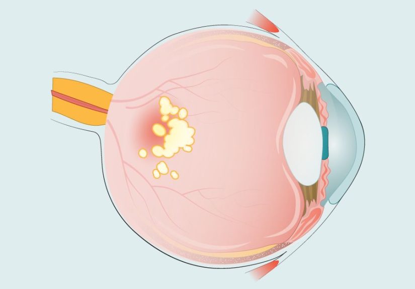
GA is an advanced form of “dry” age-related macular degeneration (AMD). In GA, patches of retinal tissue in the macula gradually thin and stop working. The macula is
the part of your retina responsible for sharp, detailed central visionthink reading, recognizing faces, and spotting the difference between “navy” and “black”
at midnight in a closet.
GA is called “geographic” because the areas of damage often look like irregular map-shapes with clear borders on retinal imaging. These damaged patches are commonly
called lesions (don’t worrythis is an eye doctor word, not a horror movie word).
GA is different from wet AMD (also called neovascular AMD). Wet AMD involves abnormal leaky blood vessels that can cause faster vision damage. Importantly,
people with GA can still develop wet AMD, either in the same eye or the other eyeso monitoring matters.
How GA Progresses: The Big Picture
GA progression usually means the atrophy lesions slowly enlarge over time. Lesions can expand outward, multiply, and sometimes merge into bigger areas. Vision changes
depend a lot on where the lesions are and whether they reach the foveathe center point of the macula that gives you the sharpest central vision.
From Early Dry AMD to GA: The Common Road
Many people don’t wake up one day with GA; it often develops after earlier stages of dry AMD. Early AMD can involve drusen (yellowish deposits under the retina) and
pigment changes. Over time, the support layers of the retinaincluding the retinal pigment epithelium and the light-sensing photoreceptorscan become damaged, leading
to areas of cell loss that define GA.
What “Progression” Means in Real Life
Clinicians often talk about GA progression in a few different ways:
- Lesion growth: the measured area of atrophy gets larger on imaging over months/years.
- Functional change: reading becomes slower, contrast looks worse, dim lighting becomes a bigger problem, or a blind spot grows.
- Location shift: lesions expand toward the center (fovea), increasing the chance of noticeable central vision impact.
A key point: GA typically progresses gradually, but the day-to-day experience can feel uneven. One month everything seems “about the same,” and then
a different month you realize you’re turning the font size up again. That doesn’t necessarily mean a sudden disease “jump”it can reflect how your brain compensates,
lighting conditions, fatigue, or small changes reaching a more noticeable spot in your visual field.
Common Milestones in GA Progression
GA doesn’t follow a perfect script, but many people recognize a few repeat “chapters”:
1) Subtle Function Changes Before Big Sharpness Loss
In earlier GA or fovea-sparing GA, central sharpness can be surprisingly decent. But other visual skills may slip firstespecially low-light vision and
contrast sensitivity. You might notice:
- More trouble seeing in dim restaurants or at dusk
- Needing brighter task lighting for reading
- Difficulty spotting curbs, steps, or low-contrast objects
- Longer “adjustment time” moving from bright to dark environments
2) Scotomas: “That Spot Where the Word Went”
As lesions enlarge, blind spots (scotomas) can become more obvious. Reading may feel like letters disappear, lines of text break apart, or the middle of a word goes
missing. Some people describe it as “a smudge,” “a blank patch,” or “someone erased the center.”
3) Fovea-Sparing vs. Fovea-Involving GA
This is one of the most useful concepts for understanding what might come next:
- Fovea-sparing GA: the center point is not yet affected. You may have a blind spot near the center, but your sharpest vision might still be present.
- Fovea-involving GA: atrophy reaches the fovea. This often leads to more noticeable central vision loss and greater difficulty with reading and face recognition.
Two people can have similarly sized lesions and very different day-to-day challenges depending on whether the fovea is involved.
4) Possible Transition Risk: Developing Wet AMD
Some people with GA develop wet AMD as well. Wet AMD can cause faster changes (like new distortion or a sudden worsening of central vision). The good news is that wet
AMD often has treatments that can help preserve vision when started promptly, so knowing the warning signs matters.
How Eye Specialists Track GA Progression
GA isn’t monitored by a single “yes/no” test. It’s tracked with a combination of eye exams, imaging, and vision function measureskind of like checking both the map
and the speedometer on a road trip.
Imaging Tools You’ll Hear About
- Optical coherence tomography (OCT): an “optical ultrasound” that shows cross-sections of retinal layers. It helps detect thinning and structural changes.
- Fundus autofluorescence (FAF): highlights patterns of retinal pigment changes and can make GA borders easier to measure.
- Color fundus photography: documents appearance over time and can support lesion tracking.
- OCT angiography (OCT-A) (sometimes): helps assess blood flow and can help detect signs that suggest wet AMD.
Vision Function Checks
Visual acuity (the eye chart) matters, but it doesn’t tell the whole storyespecially in earlier GA where reading may be hard even if chart letters look okay.
Clinicians may also consider:
- Contrast sensitivity tests (how well you see faint differences)
- Reading speed or functional reading tests
- Visual field / macular sensitivity tests (sometimes called microperimetry in specialty settings)
At-Home Monitoring: What’s Worth Doing
Your eye team may recommend at-home monitoringespecially to watch for signs of wet AMD. Options can include:
- Amsler grid checks (watching for new wavy lines or missing areas)
- Paying attention to changes in reading comfort, new distortion, or new “dark spots”
- Keeping notes about which lighting helps (so you can replicate success)
Practical tip: If you’re checking one eye at a time, do it consistently (same lighting, same distance, same time of day when possible). GA likes to play tricks when
you’re tired or the room lighting is weird.
What Can Influence How Fast GA Progresses?
GA progression varies. Some factors are fixed (thanks, biology), while others are modifiable (thanks, you).
Non-Modifiable Factors
- Age: GA risk and AMD risk increase with age.
- Genetics and family history: certain inherited risk patterns make AMD/GA more likely.
- Lesion characteristics: location (near the fovea), multiple lesions, and certain imaging patterns can be linked with different progression behavior.
Modifiable Factors (Where You Actually Get a Vote)
- Smoking: smoking is a major AMD risk factor and is strongly worth addressing.
- Cardiovascular health: blood pressure, cholesterol, and overall vascular health can matter in AMD risk discussions.
- Nutrition: diet quality matters; for certain AMD stages, specific supplements may be recommended.
- Eye care follow-up: consistent monitoring increases the chance of catching treatable changes early (like wet AMD).
This is not about “perfect lifestyle” (nobody has time for that). It’s about stacking the odds in your favorone reasonable choice at a time.
Management and Treatment: Slowing Progression and Protecting Function
For a long time, GA management focused on monitoring and adapting. That’s still importantbut in the U.S., there are now FDA-approved treatments designed to
slow GA lesion growth in many patients.
1) FDA-Approved Injections That Slow GA Lesion Growth
Two prescription eye injections are FDA-approved in the U.S. to treat GA secondary to AMD. They target parts of the immune system’s complement pathway, which research
has linked to AMD processes.
| Therapy (U.S.) | General Target | How It’s Given | Goal |
|---|---|---|---|
| pegcetacoplan | Complement C3 | Intravitreal injection (monthly or every other month, per prescribing) | Slow GA lesion growth over time |
| avacincaptad pegol | Complement C5 | Intravitreal injection (monthly, per prescribing) | Slow GA lesion growth over time |
These treatments are not a “vision reset button.” They are designed to slow how quickly the atrophy area grows. Many clinicians describe the benefit as “buying time”
potentially delaying when the lesion reaches more critical central areas for certain patients.
What These Injections Can and Can’t Do
- Can do: slow the growth rate of GA lesions on imaging for many patients in clinical trials.
- Can’t do: restore retinal cells that are already lost, or guarantee a specific amount of vision preserved.
- Reality check: the effect is measured as a reduction in lesion growth ratenot an immediate improvement in how you see the next morning.
Risks and Trade-Offs to Discuss with Your Retina Specialist
Any intravitreal injection (medicine placed into the eye) carries risks, and GA treatments have additional considerations. Topics commonly discussed include:
- Risk of developing wet AMD (neovascular AMD) in the treated eye
- Inflammation inside the eye
- Infection (endophthalmitis), which is rare but serious
- Temporary pressure increases in the eye right after injection
- Short-term effects like irritation, floaters, or small surface bleeding
Choosing treatment is a personal risk-benefit decision. Some people prioritize “doing everything possible,” while others prioritize minimizing injection burden. Many
fall somewhere in the middle: “Let’s slow it down, but let’s do it thoughtfully.”
2) Nutritional Supplements: Where AREDS2 Fits (and Where It Doesn’t)
The AREDS and AREDS2 research programs identified supplement formulas that can reduce the risk of progression from intermediate to advanced
AMD in some people. AREDS2 supplements are commonly discussed for eligible patientsespecially because the formula avoids beta-carotene (important for current/former smokers).
Important nuance: AREDS2 is not a cure for GA, and it won’t reverse existing atrophy. But if you have intermediate AMD in one or both eyes (or certain risk profiles),
your clinician may recommend AREDS2 as part of a broader strategy.
3) Low Vision Rehabilitation: The Power Tool in the Toolbox
Low vision care is not “giving up.” It’s problem-solving. A low vision specialist can help you match your actual challenges (reading mail, phone use, cooking, driving decisions)
with practical tools:
- Lighting upgrades (task lamps, glare control, strategic placement)
- Magnification (handheld magnifiers, electronic magnifiers, phone/tablet settings)
- Contrast hacks (dark cutting boards for light foods, bold labels, high-contrast clocks)
- Assistive technology (text-to-speech, audiobooks, voice assistants, large-print settings)
- Home safety (reducing fall risks, marking steps, improving hallway lighting)
Many people wait too long to ask about low vision services. If your day-to-day tasks are getting harder, it’s worth asking nownot after you’ve started negotiating with
your toaster.
A Practical “Progression Plan” You Can Use
Here’s a realistic framework (not a rigid schedule) for how people often manage GA progression with their care team:
Step 1: Establish Your Baseline
- Comprehensive dilated exam and imaging to document lesion size/location
- Discussion of whether GA is fovea-sparing or fovea-involving
- Review risk factors (smoking, family history, cardiovascular health) and a plan for what to address
Step 2: Decide on Monitoring Frequency
Monitoring frequency depends on your specific findings and risk factors. Some people need closer follow-up, especially if there are concerns about wet AMD development
or if they are receiving injections.
Step 3: Watch for “Red Flag” Symptoms
Call your eye doctor promptly if you notice:
- New wavy or distorted lines
- A sudden new dark spot or blank area
- Rapid worsening of central vision over days/weeks
- After an injection: increasing pain, worsening redness, or a major drop in vision
Step 4: Build Your Function-First Toolkit
Even if you pursue medical treatment to slow lesion growth, daily function is still the main event. A great care plan includes:
- Lighting and contrast upgrades
- Assistive technology on your phone/tablet
- Low vision rehab referrals when appropriate
- Mental health support if vision changes are affecting mood, confidence, or independence
Outlook: What to Expect Over Time
GA is generally considered a progressive condition, but the pace and the lived experience differ a lot from person to person. Many people keep useful vision for years,
especially with strong monitoring, practical adaptations, and (when appropriate) treatment to slow lesion growth. The “goal” is not perfection; it’s preserving independence,
safety, and quality of life for as long as possible.
One more hopeful point: GA research is activenew imaging tools, better functional testing, new drug strategies, and improved rehabilitation approaches continue to evolve.
Staying connected to a retina specialist and asking informed questions keeps you positioned to benefit from advances.
Experiences: What Living Through GA Progression Can Feel Like (About )
People often expect GA to announce itself with a dramatic “movie moment” (record scratch, gasp, everything goes blurry). In reality, many describe it as an accumulation
of small, annoying surprises. One of the earliest complaints is not “I can’t see,” but “Why is everything harder in dim light?” A hallway at dusk feels like it lost
30% of its contrast. A restaurant menu becomes a tiny-font prank. The irony is that in bright lighton a good dayvision can seem almost normal, which can make the
diagnosis feel confusing or even unreal.
As progression continues, reading changes in a specific way. It’s not always that letters are simply blurry; it’s that parts of words vanish. People report re-reading
the same line multiple times because their eyes “skip” missing pieces. Some switch to audiobooks or text-to-speech and feel a weird mix of relief and griefrelief
because it works, grief because it’s another reminder that the eye isn’t doing what it used to do.
A common mid-journey adjustment is learning to use peripheral vision on purpose. Many people naturally start “looking next to” what they want to seeso the image falls
on a healthier retinal area. It can feel awkward at first: you’re staring slightly off-center like you’re trying to be polite to a stranger’s forehead. But with practice,
it becomes a powerful skill, especially with coaching from low vision specialists.
Emotions are part of the progression story. Losing confidenceespecially around driving, night navigation, or recognizing facescan be as disruptive as the vision change itself.
Some people describe social situations becoming exhausting: “I’m not ignoring you, I just can’t confirm it’s you until you’re close.” Others get strategic and funny about it:
they ask people to wave, wear brighter colors, or simply announce themselves like friendly radio hosts.
For those who choose injection therapy to slow lesion growth, the experience is often described as a routine trade: a short, controlled medical discomfort in exchange for
time. People talk about planning their day around the appointment, arranging rides if their vision is temporarily off afterward, and feeling reassured by consistent monitoring.
Many also mention that the psychological benefitfeeling like there’s an active planmatters almost as much as the measurement on the scan.
The most consistent “best practice” patients share is simple: don’t wait to adapt. Better lighting, bold labels, magnification, and assistive tech are not last resorts.
They’re everyday toolslike wearing glasses, using GPS, or admitting you also can’t read the shampoo bottle in the shower. Progression may change the details, but people
often discover they can keep doing the things they lovejust with smarter setup, better support, and a little less pride about asking for help.
Conclusion
GA progression is about more than an eye scanit’s about how your vision functions in the moments that make up your day. Understanding how lesions grow, why location matters,
and how clinicians monitor change can help you make clearer decisions. Add in the newest GA-slowing treatments (for appropriate patients), consistent monitoring for wet AMD,
and early low-vision support, and you’ve got a plan built around preserving time, function, and independence.